|
Meditech Group Co.,Ltd
|

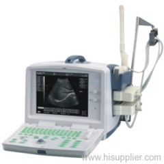
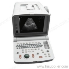
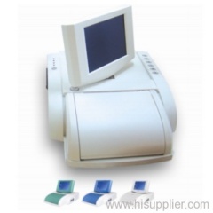
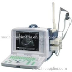
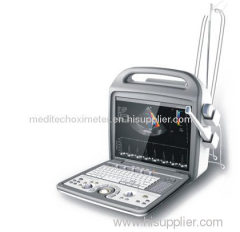
Ultrasound Scanner MD3203
| Place of Origin: | Shandong, China (Mainland) |
|
|
|
| Add to My Favorites | |
| HiSupplier Escrow |
Product Detail
Ultrasound Scanner
- Features Literature Technical Gallery Main features
- Freehand 3D and Image Optimization software packages
- Full digital with latest ultrasonic imaging techniques and embed PC
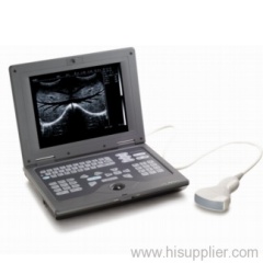
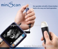
- Portable design, easy for carrying
- Back-light, silicon-gel alphameric keyboard
- Applicable for abdomen, GYN, OB, cardiac, urology and small parts
- Supporting either video or laser printer
- 5 working modes: B, B/B, B/M, M and 4B
- 10" flash-free monitor, optional 10.4" medical LCD display
- 2 probe sockets
- 8 segment TGC
- Permanent storage of 5,000 frames of ultrasound images, unlimited to
- SD card
- Peripheral ports of USB, VGA, video, DICOM3.0 and grounding
Standard configurations
- main unit
- 3.5MHz convex probe with THI
- Probe holder
- Coupling gel
Applicable Fields
Suitable for the diagnosis of Abdomen, Cardiac, Gynecology, Obstetrics, Thyroid Gland, Small Organs, Urology and so on.
Widely used for clinical examination and diagnosis. They are the ideal equipments to meet the needs of various kinds of hospitals and clinics.
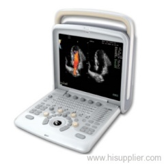
Leading Digital Technologies
Widely used for clinical examination and diagnosis. They are the ideal equipments to meet the needs of various kinds of hospitals and clinics.
Leading Digital Technologies
- DBF: Digital Beam Forming
- RDA: Real-time dynamic aperture imaging
- DRA: Dynamic real-time acoustic apodizer
- DRF: Dynamic receiving focus
- DFS: Dynamic frequency scanning, frequency range: 2.0-12.0MHz, 4 kinds of scaning frequencies
Advantages:
- Embedded computer platform is adopted in the ultrasonic master system
THI - DFC Dynamic frequency scanning
- Histogram, Sectionl drawing, Puncture guide
- Save hundreds of thousands of images and cine loop permanently
- Dynamic real-time PIP local zoom functions
- Double connectors
- Double USB ports
- Multiple kinds of OB. measurement reports, fetus physiological grades and reports and fetus growth curve
- Auto-create report systems of Gyn., small organs, cardiac, urology and other sections
- Compatible of jet printer, laser printer, video printer and video recorder
Image Processing:
- Display mode: B, 2B, 4B, B/M, M
- Image precess technologies: controllable frame correlation, Gamma correction, edge enhancement, image smoothing, image denoisiong, automatical gain adjustment, up/down, left/right and black/white conversation
- Dynamic Range: 100Db, 4 steps of switches
- Image magnification, stepless magnification, dynamic real-time PIP local zoom functions
- Cine loop: 256 frame auto/manual cine loop; multi screens cine loop (4B, 9B); auto/manual cine loop under B/M and M mode
- Image management system: the functions of pigeonholing, browsing, comparing, saving, printing and transferring images; as many as hundreds of thousands of images and thousands of cine loop could be saved; saved images could be operated by full-screen browse under slide mode.
Software Functions:
- Measurement and calculation: measure perimeter and area by distance or ellipse method; measure perimeter and area by track method; measure body surface area and volume by ellipse method. 4 measure sticks; rate measure; linear stenosis ratio, area stenosis ratio, angle measure. All calculations are automatic.
- Assist tools: puncture guide, histogram, sectional drawing
- Menu manage interface, real time online support and navigation clew system, image fore-set and one-key optimization functions.
- Auto-measure software of OB., Gyn., small organs, cardiac, urology and others:
- OB.: BPD, CRL, GS, HA, AC, HC, FL, APAD, TAD, FTA, HUMERUS, OFD, THD, TIBIA, ULNA, FI, LIMP, BBT, FBP
- Gyn.: uterus diameter, intima thickness, ovary colume, regnant ovarian follicle, length of cervix long-diameter, uterine
- Small organs: thyroid gland, hip joint
- Cardiac: AOD, LAD, IVSTd, LVIDd, AA, LAD/AOD, LVPWd, LVIDs, EF, EF SLP, CA/CE, MVCF, CO, CI, LVMWI, AVSV, FS, ACV, ET, SV, SI, LVMW, QMV
- Urology: remained urine sample, prostate, PSAD
- Presetting system for diagnosis and measurement formulas. Different formulas could be set according to different races.
- Patient cases database systems. All the data could be saved, searched and managed
- Multiple kinds of OB. measurement reports, fetus physiological grades and reports and fetus growth curve
- Auto-create report systems of Gyn., small organs, cardiac, urology and other sections
Standard Configuration:
- Main unit
- 10.4 inches SVGA high resolution non-interlaced monitor
- 256 frame auto/manual cine loop; multi screens cine loop (4B, 9B); auto/manual cine loop under B/M and M mode
- Two probe connectors
- Two USB ports
- 3.5MHz R60/R50 electronic convex transducer(2.0/5.0MHz, 128 elements)
Optional:
- 5.0MHz micro-convex probe
- 6.5MHz transvaginal probe with THI
- 7.5MHz linear probe with THI
- 7.5MHz endo-rectal probe with THI
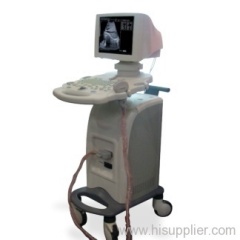
- Mobile trolley
- 10.4" medical LCD display
- Biopsy guide for convex probe
- Video printer, laser printer
- Thermal-sensitive printing paper
- SD card
Related Search
Ultrasound Scanner
Diagnostic Ultrasound Scanner
Digital Ultrasound Scanner
Veterinary Ultrasound Scanner
Ultrasound Scanner Portable
Convex Ultrasound Scanner
More>>
Find more related products in following catalogs on Hisupplier.com
Company Info
Meditech Group Co.,Ltd [China (Mainland)]
Business Type:Trading Company
City: Qingdao
Province/State: Shandong
Country/Region: China (Mainland)